Inclusions and the Micro-World: Reading Origin, Treatment, and History Under 10×
The estate ruby that could be five things
An estate client brings in a ring set with a 3.02 ct red oval in a worn yellow-gold mounting. She says it has been in her family since the 1960s and asks what you’d pay for it. Under your 10× loupe the stone is a slightly purplish red with moderate extinction, and you can see a few irregular internal features — some tiny crystals, a fingerprint-looking healed fracture near the girdle, and some faint bands of color.
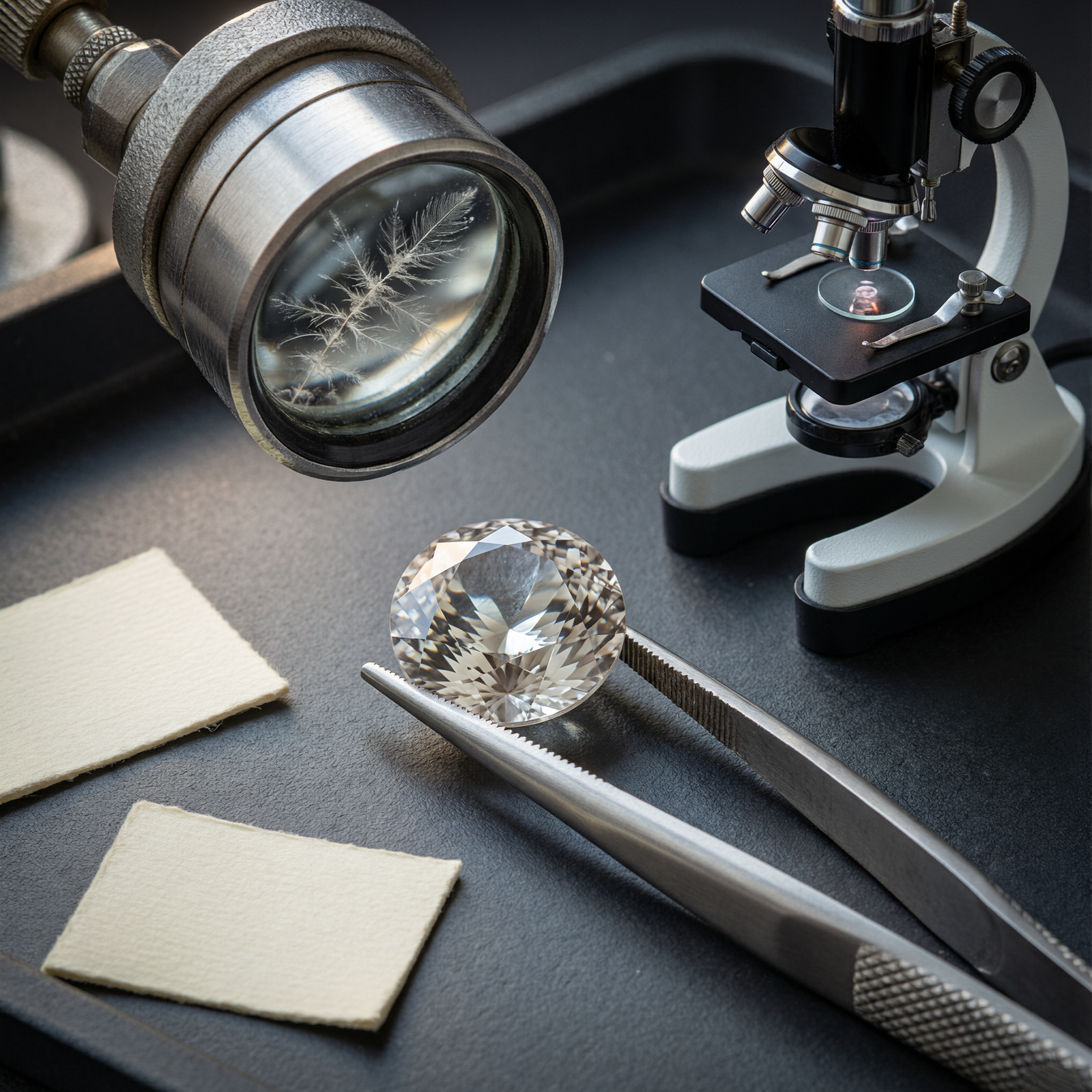
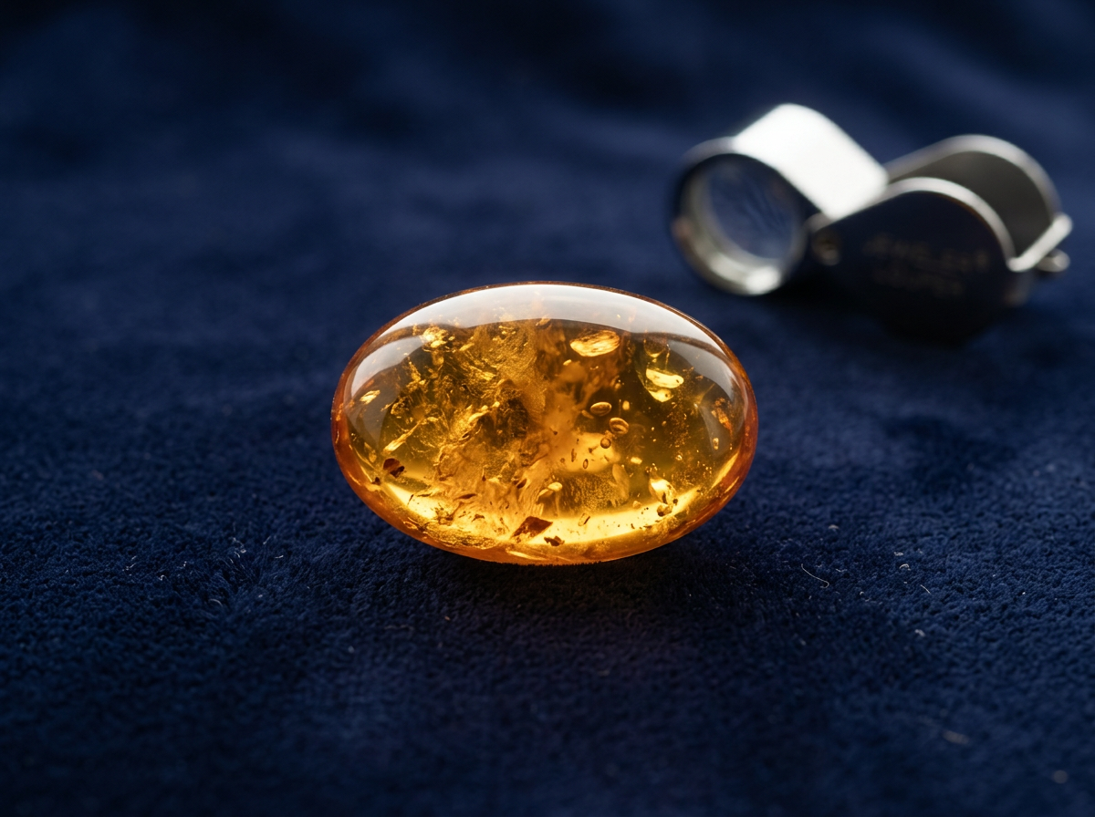
Without a microscope and spectroscopy, this stone could be five things:
1. A natural Burmese (Mogok or Mong Hsu) ruby, possibly heat-treated.
2. A natural Thai/Cambodian basalt ruby, dark and iron-rich.
3. A natural Mozambique ruby (2009 onward — unlikely if the mounting is genuinely 1960s, but mountings can be reset).
4. A Verneuil (flame-fusion) synthetic ruby from the 1960s–1970s — common in mid-century jewelry.
5. A red garnet, red spinel, red tourmaline, or doublet.
If you guess wrong you either overpay for a synthetic or garnet, or insult an estate client by under-offering on a fine Burmese stone. The right move in this situation is to say: “This is a lovely mounting. The stone is set so I’d want to examine it properly under the microscope and likely send it to a lab to confirm before I put a number on it — I’d rather give you an accurate answer than guess.”
This module explains what inclusions are, what gemologists look for under the microscope, what the famous “diagnostic” inclusions mean, what treatment inclusions look like, what synthetic inclusions look like, and — most importantly for counter work — where the line between a reasonable 10× observation ends and a professional lab opinion begins. The anchor references are the GIA Micro-World and Lab Notes columns, Eduard Gübelin and John Koivula’s Photoatlas of Inclusions in Gemstones (the field’s master reference, widely cited throughout GIA literature), and the decades of G&G treatment-detection papers.
What is an inclusion?
A strict definition of an inclusion is any material trapped inside a gem during or after its formation. The practical definition gemologists use is broader: any feature inside a gem that differs from a theoretically perfect, homogeneous crystal — including solid crystals, liquid- and gas-filled cavities, healed fractures (called “fingerprints” for their shape), growth zoning, color banding, needles and silk, cleavage and parting cracks, twin planes, exsolved crystals (crystals that formed inside the host after the host grew, as rutile silk does in corundum), and even surface-reaching features tied to growth or damage.
Think of inclusions as the gem’s internal landscape — a record of where, when, and how it grew, what heat or pressure it experienced, and whether any treatment altered it after it left the ground. Every gemological lab has a darkfield microscope — a microscope that illuminates the stone from the side against a dark background, making inclusions light up against a black field. This is the primary identification instrument in colored-stone gemology.
Inclusions are not flaws in the way a lay person might mean. They are evidence. A perfectly clean, optically flawless natural emerald is suspicious (natural emeralds almost always contain inclusions — total clarity in emerald is either synthetic, a very rare collector stone, or a clarity-enhanced stone where fractures have been filled so thoroughly they don’t show). In ruby, the presence of rutile silk is generally positive evidence of natural origin (most synthetics don’t form natural silk) and indicates the stone likely has not been heated to very high temperatures (high heat dissolves silk).
Classifying inclusions: time and type
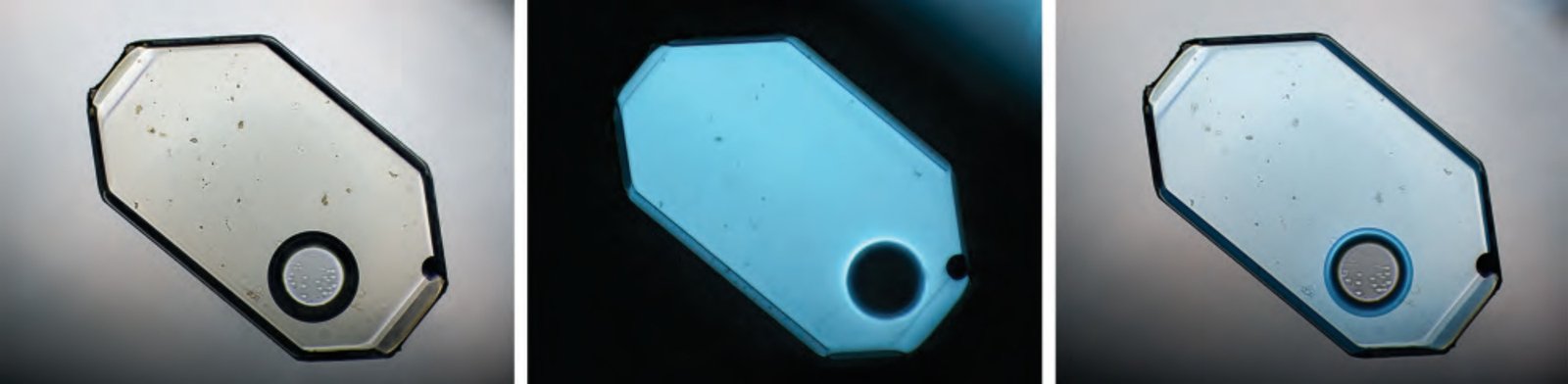
Gemologists categorize inclusions by when they formed relative to the host crystal:
- Protogenetic inclusions are pre-existing crystals from the host rock that were trapped as the gem grew around them. Calcite and dolomite crystals in marble-hosted Mogok ruby pre-date the ruby; the ruby grew around them as the marble recrystallized during metamorphism.
- Syngenetic inclusions formed at the same time as the host crystal and grew alongside it. Rutile needles (silk) in corundum are syngenetic — they exsolved from the corundum lattice as the crystal cooled, forming oriented needles along crystal directions. Three-phase inclusions in Colombian emerald formed while the emerald was growing, trapping the hydrothermal fluid and a halite crystal inside.
- Epigenetic inclusions formed after the host crystal had already grown. Healed fractures, secondary fingerprints, radiation halos around radioactive zircon crystals (zircon halos in sapphire), and fractures caused by geological stress or mining are all epigenetic.
Inclusions are also classified by how many phases (states of matter) they contain:
– Monophase: filled with liquid or gas alone.
– Two-phase (biphasic): a liquid with a gas bubble, or a solid with a gas pocket — the classic “fingerprint” healed fractures often contain two-phase fluid.
– Three-phase (triphasic): liquid + gas bubble + a solid crystal (usually a halite/salt cube) — the famous jagged three-phase inclusion in Colombian emerald is the textbook example.
Phase type plus shape plus the species of the solid crystal is the basic fingerprint for origin determination.
Famous diagnostic inclusions: what they look like and what they mean
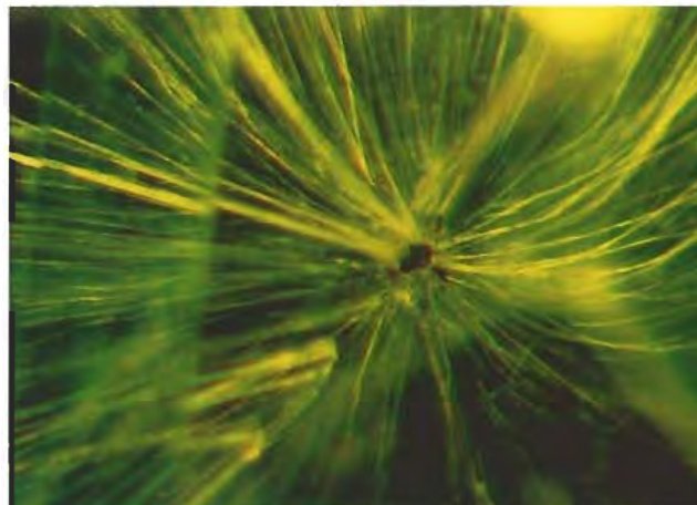
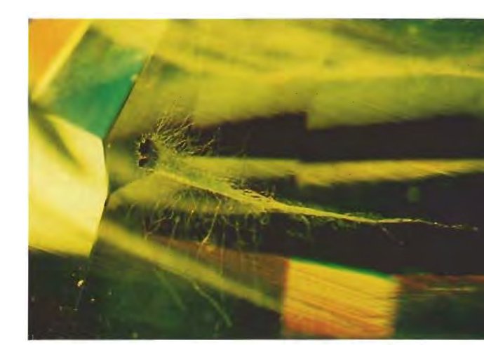
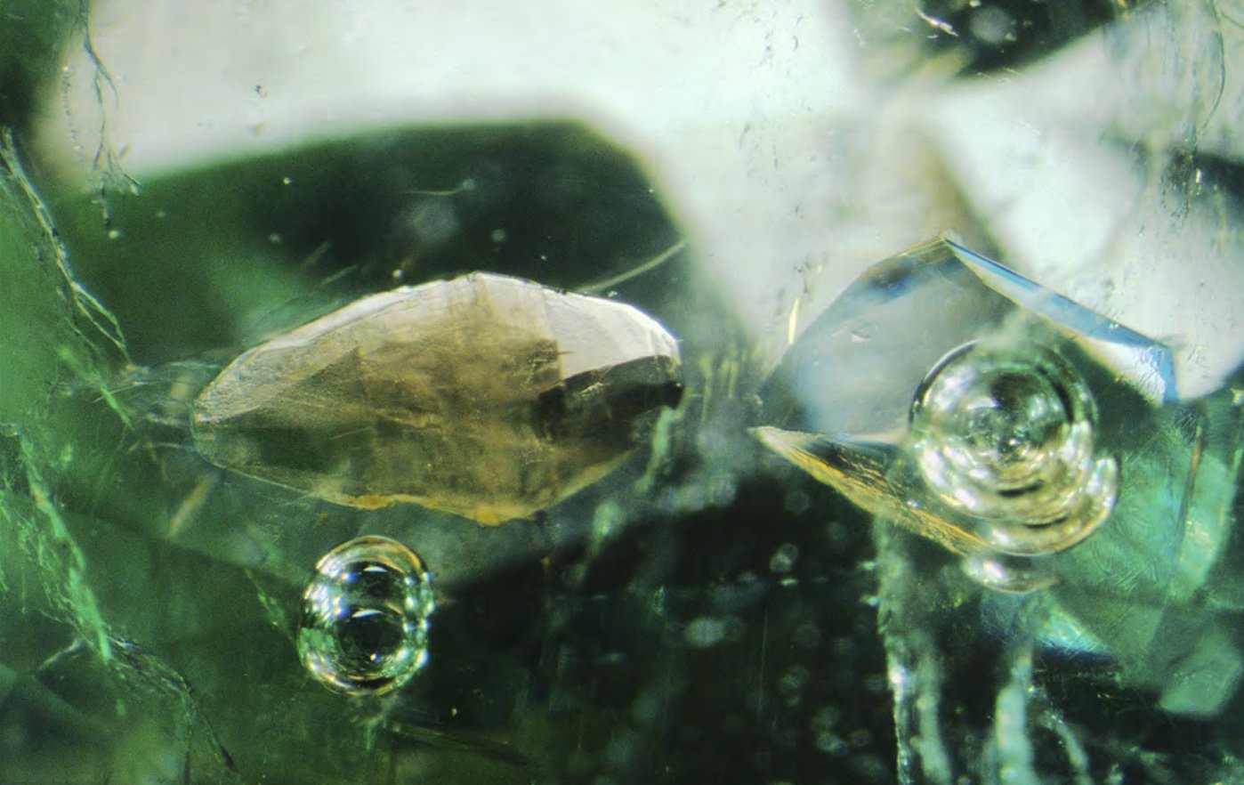
Some inclusions are so strongly associated with a particular origin or species that they act as a first-pass diagnostic. None of these are, by themselves, a guarantee — a competent lab uses multiple features together — but these are the ones every gemologist knows.
| Gem / origin | Diagnostic inclusion(s) | What they mean |
|---|---|---|
| Colombian emerald (Muzo, Chivor, Coscuez) | Three-phase jagged inclusions — a cavity containing a liquid, a gas bubble, and a tiny halite (salt) cube with a jagged/irregular shape; yellow/reddish iron-oxide staining in Muzo material; parisite, pyrite, and albite crystals. | Trapped pockets of the same hydrothermal brine the emerald grew in, precipitating a salt cube when the fluid cooled. The jagged shape and three-phase fill distinguishes Colombian from most schist-hosted emeralds. |
| Schist-hosted emerald (Zambia Kagem, Brazil Belmont/Nova Era, Sandawana Zimbabwe, Ethiopia Kenticha, Afghanistan Panjshir) | Biotite mica flakes, actinolite/tremolite needles, rectangular (rather than jagged) multiphase inclusions, atoll structures (a crystal surrounded by a ring of growth), graphite, sometimes hematite plates. | These reflect the metamorphic schist host — mica and amphibole needles are part of the schist rock the emerald formed in. Note: some schist-hosted emeralds do have multi-phase inclusions; absence of three-phase jagged inclusions is not alone proof of non-Colombian origin. |
| Kashmir sapphire | “Velvet” from submicroscopic rutile and dust-like inclusions; coarse rutile needles; pargasite crystals; zircon halos — a zircon crystal with a surrounding disc-shaped fracture caused by volume expansion from internal radiation damage over geological time. | The velvet is the reason Kashmir sapphire commands its premium — it softens the blue without making it look sleepy. Zircon halos occur in other corundum too but are classically associated with Kashmir in the literature. |
| Mogok/Burmese ruby and sapphire | Calcite and dolomite crystals (from the marble host); apatite and rutile needles; short dense rutile silk; strong angular color zoning; sometimes pyrrhotite/other sulfide inclusions. | Marble-hosted metamorphic origin; the calcite/dolomite crystals are host-rock fragments. Low iron produces bright red and strong fluorescence. |
| Thai/Cambodian basalt ruby/sapphire (Chanthaburi-Trat, Pailin) | Sparse or absent rutile silk (iron suppresses silk); reddish-brown alteration rims around crystals; pyrochlore/niobite/allanite crystals; opaque metallic inclusions (iron sulfides). | High-iron basalt host; these are the “silkless” sapphires of the basalt-field origin. |
| Mong Hsu ruby (Myanmar) | Dark blue/black core (often removed by heat treatment with flux); white/pink rim zoning; flux-healed fingerprints from routine heat treatment. | A specific Burmese deposit whose material is almost always heat-treated to remove the dark core. |
| Sri Lankan (Ceylon) sapphire (Ratnapura, Elahera) | Long, fine rutile needles (silk); prominent zircon halos; apatite and calcite crystals; distinct “fingerprint” fluid inclusions; biotite mica; long elongate negative crystals. | Classic low-iron metamorphic sapphire; the silk is why many Sri Lankan sapphires are heated (to dissolve silk for clarity or, when cooled carefully, to develop star). |
| Montana sapphire (Yogo, Rock Creek/Gem Mountain, Missouri River) | Yogo (primary deposit in a Yogo Gulch lamprophyre dike): negative crystals, distinct internal growth bands, characteristic blue-violet coloration without heat treatment; Rock Creek/Missouri River (alluvial/placer): alluvial abrasion marks on crystal surfaces, mineral inclusions from host rock, and these are commonly heat-treated after recovery. | Yogo sapphires are found in primary bedrock and have a recognized trace-element and inclusion signature; Rock Creek/Gem Mountain material is mostly alluvial and is routinely heat-treated. |
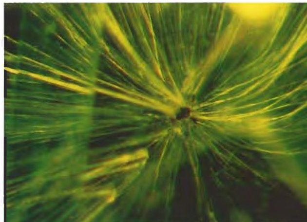
| Russian demantoid garnet (Ural Mountains) | “Horsetail” inclusions — golden-brown fibrous chrysotile/byssolite asbestos diverging in a horsetail pattern from a central chromite crystal. | The single most famous garnet inclusion. Classic Russian demantoid almost always shows horsetail inclusions; newer sources (Namibia, Madagascar, Iran, Italy) may or may not. Horsetail demantoid from the Urals commands a premium. |
| Spinel (Burma, Sri Lanka, Tajikistan) | Octahedral crystal inclusions (often other spinel crystals); iron-stained films; fingerprint fractures; apatite and calcite crystals. | Spinel is singly refractive (unlike corundum), so inclusions often look “softer” in profile. |
| Paraíba-type cuprian tourmaline (Brazil Batalha, Mozambique, Nigeria) | Trichites (hair-like fractures), fluid inclusions, copper mineral inclusions; identification is primarily spectroscopic (Cu-Mn bands), not visual. | The neon blue is colored by copper; spectroscopy (Merkel/Breeding 2009) is definitive for Paraíba-type vs. iron-colored blue/green tourmaline. |
| Ametrine (Anahi mine, Bolivia) | Distinct color zoning between purple (amethyst) and yellow (citrine) zones; typical quartz inclusions; Brazil-law twinning. | Naturally zoned quartz from hydrothermal growth; the color zones align with growth sectors that experienced different irradiation/heat histories. |
| Peridot (Myanmar, Pakistan, Arizona, China, Ethiopia) | “Lily pad” or disk-shaped healed fractures around chromite/biotite crystals; black chromite crystal inclusions; strong doubling of pavilion facets from high birefringence. | The lily-pad inclusions are characteristic and a quick ID aid under 10×. |
Treatment inclusions: what treated stones look like under 10×
Treatments leave telltale evidence. Sometimes you can see it at 10× with a loupe; usually you need a microscope. What to look for, summarized:
| Treatment | Visible features (10× to microscope) |
|---|---|
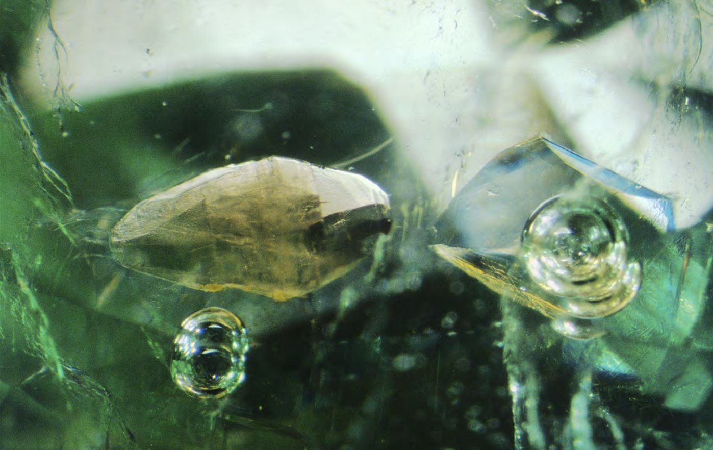
| Heat-treated corundum (most commercial ruby/sapphire) | Dissolved/altered/coarsened rutile silk; “snowflake” inclusions around altered crystals; circular discoid fractures surrounding crystal inclusions (from differential thermal expansion); pitted girdle surface if overheated; sintered areas around cavities. Heat treatment without filling does not add foreign material — you are looking for altered inclusions, not added substance. |
| Surface-diffused blue sapphire (older Ti-diffusion, less common today) | Blue color concentrated at facet edges and girdle; when a stone is repolished after diffusion, color can be polished through on one or two facets. |
| Beryllium-diffused sapphire | Cannot reliably be seen by eye — detection requires chemical analysis (LIBS/LA-ICP-MS or photoluminescence); surface features can include a frosted rim after treatment, and colors are often atypically bright, orangey-pinks or yellows. Do not attempt to call Be-diffusion by eye. |
| Lead-glass-filled ruby | Flash effect (bright blue or orange flashes at filled fractures when tilted in reflected light); large gas bubbles trapped in the glass; flattened bubbles at the glass/corundum interface; uneven surface luster on filled fractures; filler can show a different gloss than corundum at facet junctions. The four detection features in GIA-CS-06 are: flash effect, large gas bubbles, flattened gas bubbles, and durability warning. |
| Oiled/resin-filled emerald | Flattened gas bubbles in surface-reaching fissures; filler may show a yellowish or bluish flash in reflected light; dried filler appears chalky in older stones; filler drips or pooling on the surface of poorly filled stones; in UV, some resins fluoresce. Filler is usually only visible under magnification in the fissures. |
| Irradiated blue topaz | No visible inclusions from the irradiation itself — detection is by spectroscopy or is not necessary (all commercial blue topaz is irradiated, and disclosure is color-process disclosure). |
| Coated tanzanite / mystic topaz | Bubbles in coating; coating thickness variations visible on facet edges; color scrapes off the girdle if abraded (do not test on client-owned stones); uncoated pavillon/culet area (if coating is applied to pavilion only). |
| Dyed stones (dyed lapis, C-jade, dyed turquoise, dyed chalcedony, dyed howlite “turquoise”) | Dye concentration along fractures and grain boundaries; uneven color; swab test with acetone may pick up dye (do not test on client stones without permission); dyed lapis may show dye concentrations around pyrite grains. |
| Sugar/acid-treated Andamooka matrix opal (Brown 1991) | Darkened bodycolor from carbonized sugar in pore spaces; patchy darkening; surface-concentrated darkening. |
| Bleached/polymer-impregnated jade (B-jade) | Cannot be reliably identified by eye; infrared spectroscopy detects polymer resin; visible features include unusually “loose” texture, pitted surface, and a duller luster, but these are not definitive. |
[MEDIA: link-out | C17-M06-L1]
Reference: How to Classify a Lead Glass–Filled Ruby — GIA (GIA-CS-06, already in the video library)
Demonstrates flash effect, large gas bubbles, flattened bubbles, and the durability warning. 5 min.
https://www.youtube.com/watch?v=evrQv-HNrI0
Synthetic signatures: what man-made stones look like under magnification
Synthetics are covered in depth in M08, but the classic 10× signatures are worth knowing here — because if you see curved striae in a “ruby,” you don’t need a lab to tell you it is not natural.
| Growth method | Species grown | Key visual features |
|---|---|---|
| Verneuil (flame-fusion) | Ruby, sapphire, blue/white spinel simulants, rutile, strontium titanate | Curved growth striae (curved color bands) — single most important diagnostic feature; these are the growth lines from the rotating boule and never appear in natural corundum (natural color bands are straight/angular); spherical gas bubbles (often large, isolated and dark, not fingerprint-like in fluid; bubbles in glass fill look different from bubbles in Verneuil synthetics because glass fill follows fractures, while Verneuil bubbles are distributed through the boule); sharp, pinpoint Plato lines in certain orientations. |
| Czochralski (pulled) | Ruby, sapphire, alexandrite, YAG, GGG | Curved striae can be present; gas bubbles; sometimes very clean internally; used for many alexandrite synthetics and laser ruby. |
| Flux-melt | Ruby, sapphire, emerald, alexandrite, spinel | Wispy/veil-like flux inclusions (often a “chevron” or “lace” pattern); platinum crystal platelets from platinum crucibles; “feather” flux that looks too regular or whitish; no natural mineral inclusions of the host-rock type (no calcite/biotite/actinolite — the kind of inclusions natural gems carry from their host rock). Chatham, Gilson, Kyocera and others use flux processes. |
| Hydrothermal | Emerald (Biron, Pool, Russian, Chinese hydrothermal), quartz, synthetic ruby/sapphire less commonly | Chevron / zig-zag growth patterns (the single most quoted feature of hydrothermal synthetics); nail-head spicules (in hydrothermal synthetic emeralds especially); phenakite crystals; metal platelets (gold, copper) from the autoclave; tapered two-phase fluid inclusions; seed plate visible in some stones (a flat line marking the seed crystal). |
| Synthetic opal (Gilson, Kyocera) | White/black opal imitation | Columnar “lizard skin” structure viewed perpendicular to the play-of-color surface; array of spheres is unnaturally regular in size and arrangement compared to natural opal; often no host-rock matrix (boulder ironstone host = natural Australian by definition). |
| Synthetic moissanite (SiC) | Diamond simulant | Doubled facet junctions (very high birefringence); fine white needle inclusions; strings of bubbles; detected instantly with a diamond tester combination (moissanite has different thermal/electrical conductivity). Covered in C16, not here in depth. |
The single counter rule: if you see curved growth striae in a red or blue stone, it is Verneuil synthetic corundum until proven otherwise. Refer to a lab to confirm but treat as synthetic for valuation purposes.
How labs actually do origin determination
If inclusions alone were enough, origin reports would be quick and cheap. In practice, a modern GIA colored-stone origin report combines at least three lines of evidence:
1. Microscopic inclusions — the internal features described above.
2. Trace-element chemistry — LA-ICP-MS (laser ablation inductively-coupled plasma mass spectrometry) measures the concentrations of trace elements (Mg, Ti, V, Cr, Fe, Ga, etc.) to a parts-per-million precision. Different mines have different chemical fingerprints even when they produce the same species; Madagascar sapphires, for instance, can have different trace-element signatures from Sri Lankan sapphires even when the inclusions look similar.
3. Spectroscopy — FTIR (Fourier-transform infrared) detects treatments such as heating and filling; UV-Vis-NIR spectroscopy measures the absorption spectrum (which tells you which elements are causing the color and can distinguish natural vs treated); Raman spectroscopy identifies individual inclusion minerals (e.g., confirming that a crystal inside a Colombian emerald is parisite rather than apatite).
4. Photomicrography and comparison to a reference library built from the field-gemology trips described in M03. The direct-from-mine samples Pardieu, Vertriest and others collect at the jig become the reference set.
Because origin determination combines multiple lines of evidence and mines in the same region can produce overlapping chemistry, origin reports are expert judgments, not mathematical certainties. When the lab cannot reach a confident conclusion on a particular stone it will issue an “inconclusive” report rather than guess. This is a feature, not a failure — it is better for a lab to say “we cannot determine origin” than to issue a wrong origin.
[MEDIA: link-out | C17-M06-L2]
Reference: A Gemstone’s Journey through the GIA Laboratory (GIA-CS-01) darkfield microscope segment (~25–30 min mark) shows Nathan Renfro demonstrating microscopy and spectroscopy.
https://www.youtube.com/watch?v=P5J1a3_JcTM
Loupe discipline and when to refer to a lab
Proper 10× loupe technique takes an hour to learn and years to master. The basics:
1. Hold the loupe close to your eye (your eyelashes should nearly touch the lens).
2. Bring the stone up to the loupe from a couple of inches away, holding it with tweezers.
3. Tilt and rock the stone under an overhead light source so light travels through the pavilion and reflects off inclusions.
4. Look at the table first, then tilt to examine every facet, then check the girdle and culet carefully (coating damage, chips, and glass fill are often easiest to see at the girdle).
5. Darkfield illumination (a light source from the side against a dark background) is dramatically more revealing than direct overhead light; every store should have one.
Refer to a gemologist or lab, rather than guessing, when:
– You see curved striae (possible Verneuil synthetic).
– You see flash effect or gas bubbles in fractures (possible lead-glass fill).
– You see bubbles in coating, or coating at facet edges.
– You see unusual flux/chevron inclusions (possible flux or hydrothermal synthetic).
– The client asks for a written origin determination or natural/synthetic guarantee.
– The stone is a high-value estate piece where identity, origin, or treatment affects price materially.
– You are unsure. “I don’t know yet — let me have our gemologist look at it” is always a professional answer. It is never professional to guess.
On the floor: applying it this week
- Monday (10 minutes): If your store has a darkfield loupe or a microscope, ask the gemologist to show you a known three-phase inclusion in a Colombian emerald and rutile silk in a sapphire. Seeing these once anchors the module.
- Tuesday (10 minutes): Practice proper loupe technique on five stones in your case — hold the loupe close, bring the stone to it, rock the stone. Do this until it feels natural.
- Wednesday (practice): When examining a customer or estate stone, discipline yourself to say “I can see some interesting features under the loupe — I’d like to put this under the microscope/send it to our gemologist before I give you a definitive answer” if you see anything you cannot identify confidently.
- Thursday: Read one GIA Micro-World or Lab Notes column entry from the bibliography — any one. The photomicrographs are the fastest way to build an internal library of what inclusions look like.
- Friday (5 minutes): Watch GIA-CS-06 (the 5-minute lead-glass-filled ruby detection video) if you haven’t yet — being able to spot flash effect in a filled ruby is one of the highest-value counter skills you can develop.
Objections, mistakes and edge cases
| Situation | The trap | Better move |
|---|---|---|
| Client brings in an estate “ruby” | Guessing it is natural because the mounting is old | Curved-striae Verneuil synthetics have been around since the early 1900s; many 20th-century mountings are set with synthetics. Loupe it; refer for a report before you put a number on it. |
| Client wants to know the origin of an unreported sapphire | “It looks like Ceylon to me” based on color alone | Origin cannot be called by color. Inclusions + chemistry + spectroscopy are needed. Tell the client you can send it to the lab for an origin report. |
| Client says “this emerald is perfectly clean — so it must be the best” | Agreeing that clean = finest | Natural emerald almost always contains inclusions (jardin). Very clean emeralds are either synthetic, heavily filled, or rare collector material; a truly eye-clean natural emerald should be examined closely. The jardin is part of emerald’s identity — many collectors prefer it. |
| You see flash effect in a fracture in a red stone | Declaring it “lead-glass-filled ruby” immediately | Flash effect can occasionally be seen in other fill situations; large and flattened gas bubbles plus durability context are required for a confident call. But if you see flash effect, treat the stone as suspect and refer. |
| Client has a “star sapphire” with a perfectly sharp, dead-centered star that doesn’t move | Assuming all stars are natural | Diffusion-treated synthetic stars (and engraved-star doublets per McClure/Koivula 2001) produce unnaturally perfect stars that don’t move naturally with the light. Refer. |
| You see no inclusions at all in a colored stone | Concluding it is synthetic | A small percentage of natural colored stones (some aquamarine, morganite, garnet, tanzanite) can be loupe-clean. Absence of inclusions is not by itself proof of synthesis. Refer. |
| Client asks you to identify a stone in a mounting while they wait | Guessing by color and cut | Color is not identity. Use RI, SG, spectroscopy (if you have the tools); otherwise say “I can give you my best guess but I can’t confirm here — let me send it to the lab.” |
| Edge case: lead-glass filled ruby sold as “ruby, filled composite ruby” | Treating it as equivalent to natural ruby | Disclosure is required (M09); filled ruby is a composite stone with different durability and value than solid natural ruby. |
Self-check
- What does a darkfield microscope do that makes inclusions visible?
- Name the three time categories of inclusions (when they formed relative to the host).
- What is a three-phase inclusion, and in which origin of emerald is the jagged three-phase inclusion considered classic?
- What inclusion is the most famous diagnostic for Russian demantoid garnet?
- What does “jardin” mean in emerald?
- When a corundum has been heated, what visible changes to inclusions might you see under a microscope?
- What are the four detection features GIA lists for lead-glass-filled ruby (GIA-CS-06)?
- What single feature, if you see it under 10×, should make you strongly suspect a Verneuil (flame-fusion) synthetic ruby or sapphire?
- What two tools beyond the microscope does a modern lab use for colored-stone origin determination?
- When should you stop looking at a stone under a loupe and refer to a gemologist/lab instead of giving a counter opinion?
Go deeper
- GIA Photomicrography / Micro-World gallery — https://www.gia.edu/photomicrography — hundreds of inclusion photographs; browse for 10 minutes to build your visual library.
- How to Classify a Lead Glass–Filled Ruby (GIA-CS-06) — https://www.youtube.com/watch?v=evrQv-HNrI0 — 5 min, already verified.
- A Gemstone’s Journey through the GIA Laboratory (GIA-CS-01) — darkfield/spectroscopy segment at ~25–30 min — https://www.youtube.com/watch?v=P5J1a3_JcTM.
- Gübelin E.J., Koivula J.I. (2005) Photoatlas of Inclusions in Gemstones, Vols. 1–3. The master reference work (not in GIA’s PDF reading list; this is a textbook cited throughout GIA literature — not freely available online, but every gemologist’s desk has a set).
- Koivula J.I., Kammerling R.C. et al. — ongoing “Gem News” and “Micro-World” columns in G&G across the decades; search gia.edu for “micro-world” or “lab notes” and the species of interest.
- Saeseaw S. et al. (2014) Geographic Origin Determination of Emerald and related origin-determination papers (see bib) for how labs combine inclusions + chemistry.
- McClure S.F. (2006) Lead-glass-filled rubies papers — G&G (multiple entries) for treatment detection.
- Schmetzer K. (1987, 2015) synthetic-opal and double-star papers — cited for synthetic-opal structure and dual stars.
- Emmett J.L. et al. (2003) Beryllium Diffusion of Ruby and Sapphire — G&G (cited) for beryllium detection chemistry.